Last updated on 2019-09-30 by Xiang-Jun Lu <xiangjun@x3dna.org>.
The block schematics were created with DSSR and
rendered using PyMOL.
- PDB-id
- 5T01
- Class
- transcription regulator-DNA
- Method
- X-ray (1.89 Å)
- Summary
- Human c-jun DNA binding domain homodimer in complex with methylated DNA
List of 1 5mC-amino acid contact:
-
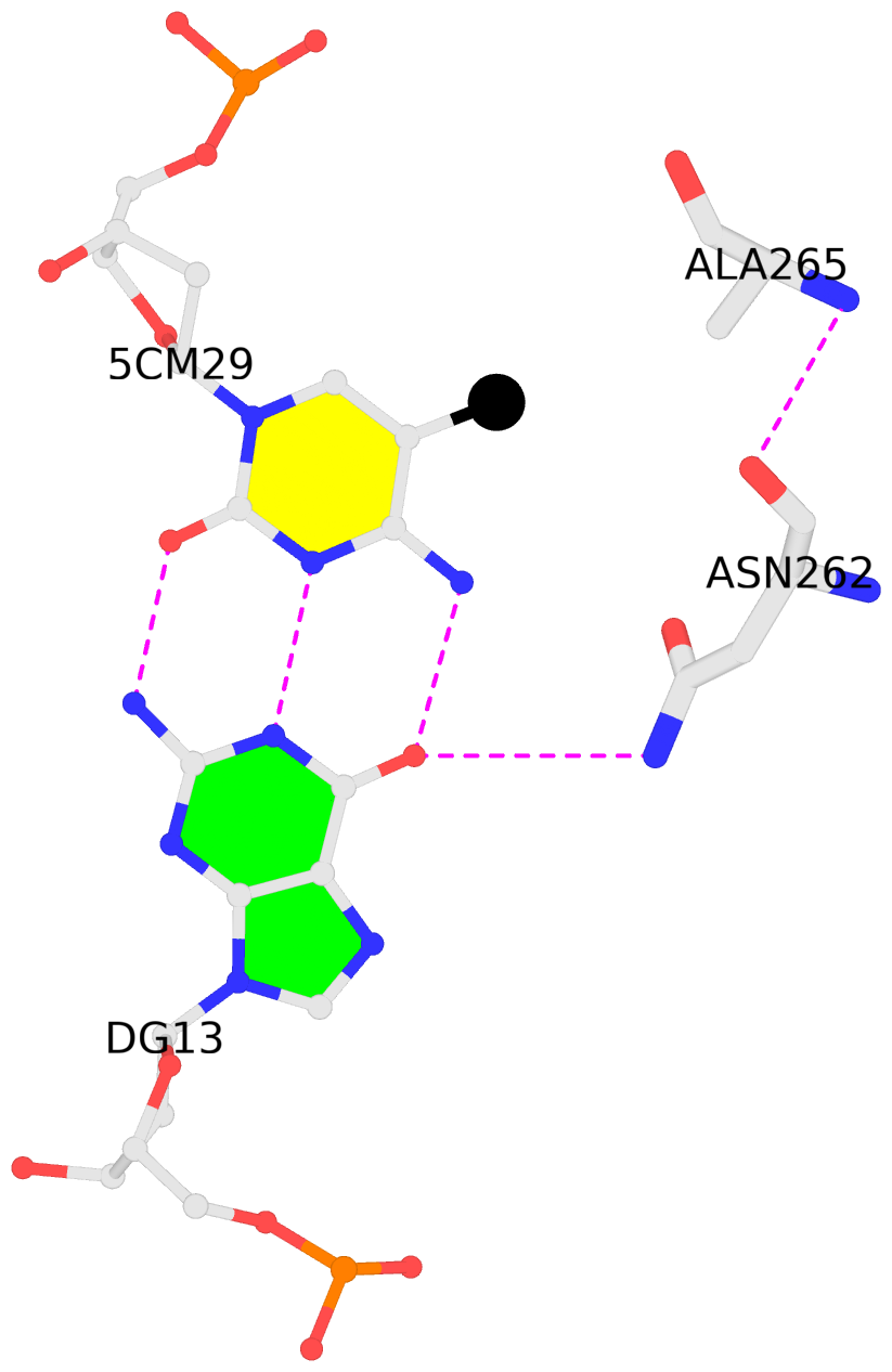
D.5CM29: hydrophobic-with-A.ALA265 is-WC-paired is-in-duplex [-]:CGT/AcG
direct SNAP output · DNAproDB 2.0
- Reference
- Hong, S., Wang, D., Horton, J.R., Zhang, X., Speck, S.H., Blumenthal, R.M., Cheng, X.: (2017) "Methyl-dependent and spatial-specific DNA recognition by the orthologous transcription factors human AP-1 and Epstein-Barr virus Zta." Nucleic Acids Res., 45, 2503-2515.
- Abstract
- Activator protein 1 (AP-1) is a transcription factor that recognizes two versions of a 7-base pair response element, either 5΄- GAG CA-3΄ or 5΄- GAG CA-3΄ (where M = 5-methylcytosine). These two elements share the feature that 5-methylcytosine and thymine both have a methyl group in the same position, 5-carbon of the pyrimidine, so each of them has two methyl groups at nucleotide positions 1 and 5 from the 5΄ end, resulting in four methyl groups symmetrically positioned in duplex DNA. Epstein-Barr Virus Zta is a key transcriptional regulator of the viral lytic cycle that is homologous to AP-1. Zta recognizes several methylated Zta-response elements, including meZRE1 (5΄- GAG C A-3΄) and meZRE2 (5΄- GAG G A-3΄), where a methylated cytosine occupies one of the inner thymine residues corresponding to the AP-1 element, resulting in the four spatially equivalent methyl groups. Here, we study how AP-1 and Zta recognize these methyl groups within their cognate response elements. These methyl groups are in van der Waals contact with a conserved di-alanine in AP-1 dimer (Ala265 and Ala266 in Jun), or with the corresponding Zta residues Ala185 and Ser186 (via its side chain carbon Cβ atom). Furthermore, the two ZRE elements differ at base pair 6 (C:G versus G:C), forming a pseudo-symmetric sequence (meZRE1) or an asymmetric sequence (meZRE2). In vitro DNA binding assays suggest that Zta has high affinity for all four sequences examined, whereas AP-1 has considerably reduced affinity for the asymmetric sequence (meZRE2). We ascribe this difference to Zta Ser186 (a unique residue for Zta) whose side chain hydroxyl oxygen atom interacts with the two half sites differently, whereas the corresponding Ala266 of AP-1 Jun protein lacks such flexibility. Our analyses demonstrate a novel mechanism of 5mC/T recognition in a methylation-dependent, spatial and sequence-specific approach by basic leucine-zipper transcriptional factors.
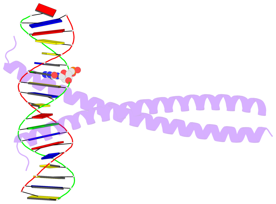
- The 5-methylcytosine group (PDB ligand '5CM') is shown in space-filling model,
with the methyl-carbon atom in black.
- Watson-Crick base pairs are represented as long rectangular blocks with the
minor-groove edge in black. Color code: A-T red, C-G yellow, G-C green, T-A blue.
- Protein is shown as cartoon in purple. DNA backbones are shown ribbon, colored code
by chain identifier.
- The block schematics were created with 3DNA-DSSR,
and images were rendered using PyMOL.
- Download the PyMOL session file corresponding to the top-left
image in the following panel.
- The contacts include paired nucleotides (mostly a G in G-C pairing), and
amino-acids within a 4.5-A distance cutoff to the base atoms of 5mC.
- The structure is oriented in the 'standard' base reference frame of 5mC, allowing for easy comparison
and direct superimposition between entries.
- The black sphere (•) denotes the 5-methyl carbon atom in 5mC.
 |
No. 1 D.5CM29: download PDB file
for the 5mC entry
hydrophobic-with-A.ALA265 is-WC-paired is-in-duplex [-]:CGT/AcG
|


