Last updated on 2019-09-30 by Xiang-Jun Lu <xiangjun@x3dna.org>.
The block schematics were created with DSSR and
rendered using PyMOL.
- PDB-id
- 5CPK
- Class
- structural protein-DNA
- Method
- X-ray (2.63 Å)
- Summary
- Nucleosome containing methylated sat2l DNA
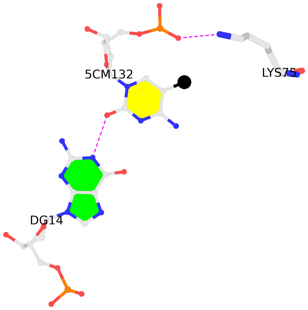
List of 1 5mC-amino acid contact:
-
I.5CM132: other-contacts is-WC-paired is-in-duplex [+]:TcG/cGA
direct SNAP output · DNAproDB 2.0
- Reference
- Osakabe, A., Adachi, F., Arimura, Y., Maehara, K., Ohkawa, Y., Kurumizaka, H.: (2015) "Influence of DNA methylation on positioning and DNA flexibility of nucleosomes with pericentric satellite DNA." Open Biology, 5
- Abstract
- DNA methylation occurs on CpG sites and is important to form pericentric heterochromatin domains. The satellite 2 sequence, containing seven CpG sites, is located in the pericentric region of human chromosome 1 and is highly methylated in normal cells. In contrast, the satellite 2 region is reportedly hypomethylated in cancer cells, suggesting that the methylation status may affect the chromatin structure around the pericentric regions in tumours. In this study, we mapped the nucleosome positioning on the satellite 2 sequence in vitro and found that DNA methylation modestly affects the distribution of the nucleosome positioning. The micrococcal nuclease assay revealed that the DNA end flexibility of the nucleosomes changes, depending on the DNA methylation status. However, the structures and thermal stabilities of the nucleosomes are unaffected by DNA methylation. These findings provide new information to understand how DNA methylation functions in regulating pericentric heterochromatin formation and maintenance in normal and malignant cells.
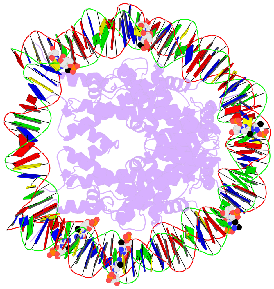
- The 5-methylcytosine group (PDB ligand '5CM') is shown in space-filling model,
with the methyl-carbon atom in black.
- Watson-Crick base pairs are represented as long rectangular blocks with the
minor-groove edge in black. Color code: A-T red, C-G yellow, G-C green, T-A blue.
- Protein is shown as cartoon in purple. DNA backbones are shown ribbon, colored code
by chain identifier.
- The block schematics were created with 3DNA-DSSR,
and images were rendered using PyMOL.
- Download the PyMOL session file corresponding to the top-left
image in the following panel.
- The contacts include paired nucleotides (mostly a G in G-C pairing), and
amino-acids within a 4.5-A distance cutoff to the base atoms of 5mC.
- The structure is oriented in the 'standard' base reference frame of 5mC, allowing for easy comparison
and direct superimposition between entries.
- The black sphere (•) denotes the 5-methyl carbon atom in 5mC.
 |
No. 1 I.5CM132: download PDB file
for the 5mC entry
other-contacts is-WC-paired is-in-duplex [+]:TcG/cGA
|


