Last updated on 2019-09-30 by Xiang-Jun Lu <xiangjun@x3dna.org>.
The block schematics were created with DSSR and
rendered using PyMOL.
- PDB-id
- 4R2R
- Class
- DNA binding protein-DNA
- Method
- X-ray (2.09 Å)
- Summary
- Wilms tumor protein (wt1) zinc fingers in complex with carboxylated DNA
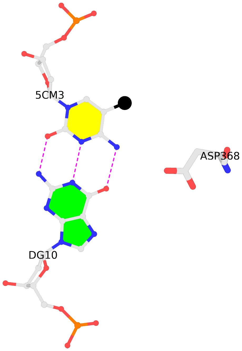
List of 1 5mC-amino acid contact:
-
C.5CM3: other-contacts is-WC-paired is-in-duplex [-]:cGT/AcG
direct SNAP output · DNAproDB 2.0
- Reference
- Hashimoto, H., Olanrewaju, Y.O., Zheng, Y., Wilson, G.G., Zhang, X., Cheng, X.: (2014) "Wilms tumor protein recognizes 5-carboxylcytosine within a specific DNA sequence." Genes Dev., 28, 2304-2313.
- Abstract
- In mammalian DNA, cytosine occurs in several chemical forms, including unmodified cytosine (C), 5-methylcytosine (5 mC), 5-hydroxymethylcytosine (5 hmC), 5-formylcytosine (5 fC), and 5-carboxylcytosine (5 caC). 5 mC is a major epigenetic signal that acts to regulate gene expression. 5 hmC, 5 fC, and 5 caC are oxidized derivatives that might also act as distinct epigenetic signals. We investigated the response of the zinc finger DNA-binding domains of transcription factors early growth response protein 1 (Egr1) and Wilms tumor protein 1 (WT1) to different forms of modified cytosine within their recognition sequence, 5'-GCG(T/G)GGGCG-3'. Both displayed high affinity for the sequence when C or 5 mC was present and much reduced affinity when 5 hmC or 5 fC was present, indicating that they differentiate primarily oxidized C from unoxidized C, rather than methylated C from unmethylated C. 5 caC affected the two proteins differently, abolishing binding by Egr1 but not by WT1. We ascribe this difference to electrostatic interactions in the binding sites. In Egr1, a negatively charged glutamate conflicts with the negatively charged carboxylate of 5 caC, whereas the corresponding glutamine of WT1 interacts with this group favorably. Our analyses shows that zinc finger proteins (and their splice variants) can respond in modulated ways to alternative modifications within their binding sequence.
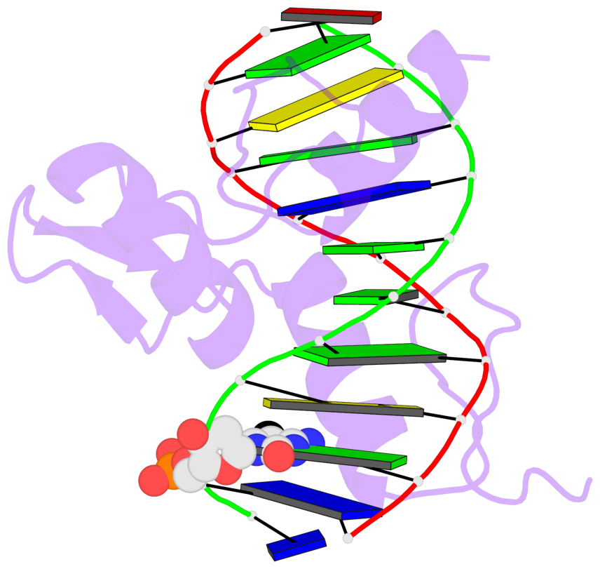
- The 5-methylcytosine group (PDB ligand '5CM') is shown in space-filling model,
with the methyl-carbon atom in black.
- Watson-Crick base pairs are represented as long rectangular blocks with the
minor-groove edge in black. Color code: A-T red, C-G yellow, G-C green, T-A blue.
- Protein is shown as cartoon in purple. DNA backbones are shown ribbon, colored code
by chain identifier.
- The block schematics were created with 3DNA-DSSR,
and images were rendered using PyMOL.
- Download the PyMOL session file corresponding to the top-left
image in the following panel.
- The contacts include paired nucleotides (mostly a G in G-C pairing), and
amino-acids within a 4.5-A distance cutoff to the base atoms of 5mC.
- The structure is oriented in the 'standard' base reference frame of 5mC, allowing for easy comparison
and direct superimposition between entries.
- The black sphere (•) denotes the 5-methyl carbon atom in 5mC.
 |
No. 1 C.5CM3: download PDB file
for the 5mC entry
other-contacts is-WC-paired is-in-duplex [-]:cGT/AcG
|


