Last updated on 2019-09-30 by Xiang-Jun Lu <xiangjun@x3dna.org>.
The block schematics were created with DSSR and
rendered using PyMOL.
- PDB-id
- 4GJR
- Class
- transcription-DNA
- Method
- X-ray (1.85 Å)
- Summary
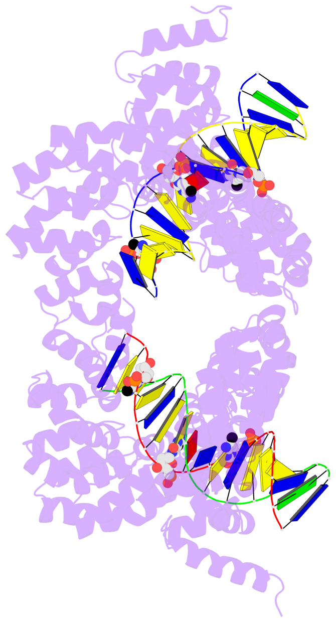
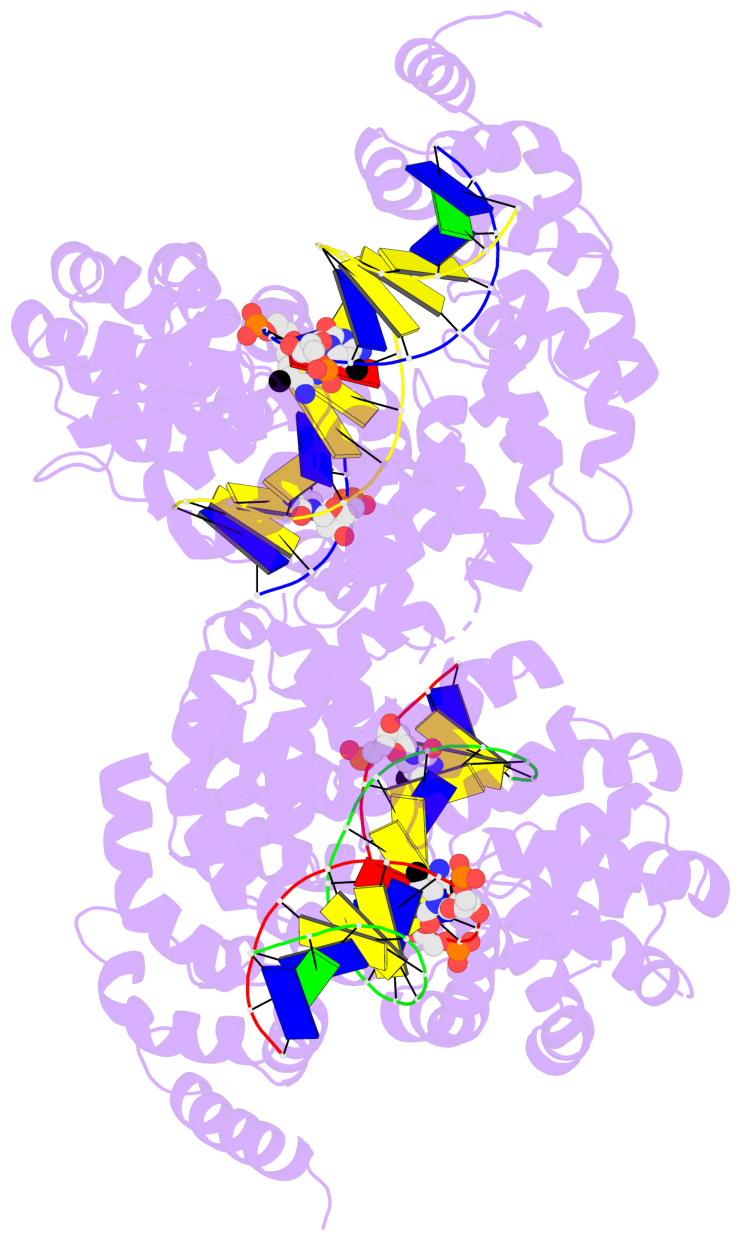
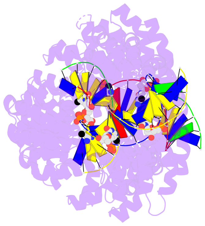
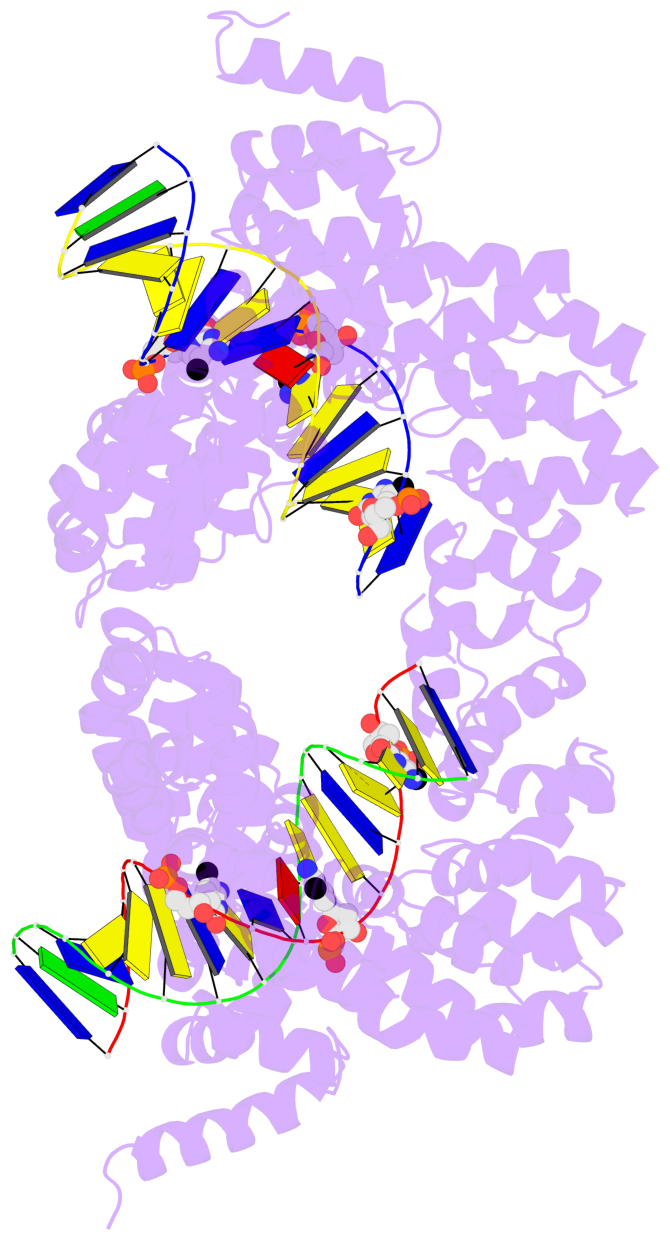
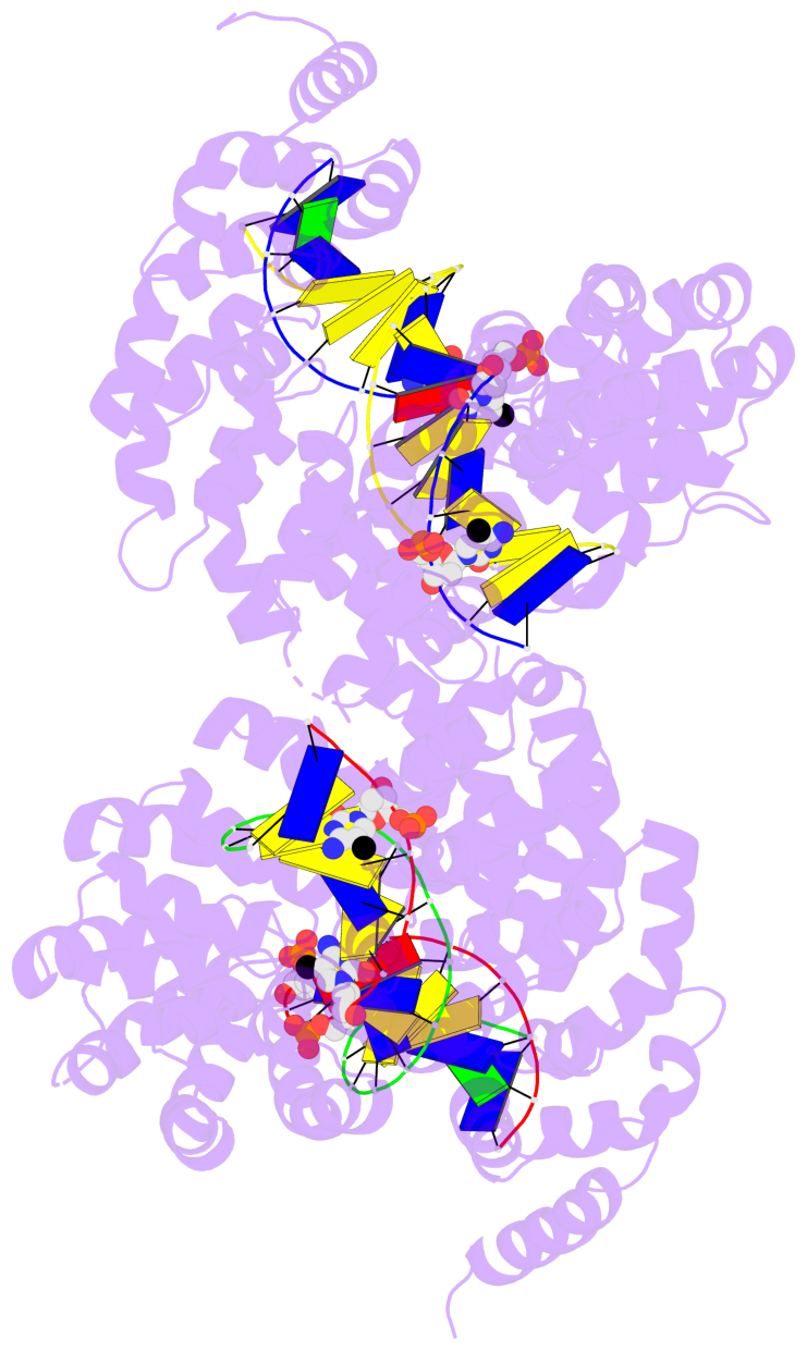
- Crystal structure of the tal effector dhax3 bound to methylated dsDNA
List of 6 5mC-amino acid contacts:
-
I.5CM8: hydrophobic-with-A.GLY437 hydrophobic-with-A.GLY438 is-WC-paired is-in-duplex [+]:TcT/AGA
-
I.5CM11: stacking-with-A.ASP573 is-WC-paired is-in-duplex [+]:AcC/GGT
-
I.5CM15: hydrophobic-with-A.GLY675 hydrophobic-with-A.GLY676 is-WC-paired is-in-duplex [+]:CcC/GGG
-
G.5CM8: hydrophobic-with-B.GLY437 hydrophobic-with-B.GLY471 hydrophobic-with-B.GLY472 is-WC-paired is-in-duplex [+]:TcT/AGA
-
G.5CM11: stacking-with-B.ASP573 is-WC-paired is-in-duplex [+]:AcC/GGT
-
G.5CM15: hydrophobic-with-B.GLY675 hydrophobic-with-B.GLY676 is-WC-paired is-in-duplex [+]:CcC/GGG
direct SNAP output · DNAproDB 2.0
- Reference
- Yan, N., Deng, D., Yan, C.Y., Yin, P., Pan, X.J., Shi, Y.G.: "Crystal structure of a protein complex." To be Published
- Abstract
-
- The 5-methylcytosine group (PDB ligand '5CM') is shown in space-filling model,
with the methyl-carbon atom in black.
- Watson-Crick base pairs are represented as long rectangular blocks with the
minor-groove edge in black. Color code: A-T red, C-G yellow, G-C green, T-A blue.
- Protein is shown as cartoon in purple. DNA backbones are shown ribbon, colored code
by chain identifier.
- The block schematics were created with 3DNA-DSSR,
and images were rendered using PyMOL.
- Download the PyMOL session file corresponding to the top-left
image in the following panel.
- The contacts include paired nucleotides (mostly a G in G-C pairing), and
amino-acids within a 4.5-A distance cutoff to the base atoms of 5mC.
- The structure is oriented in the 'standard' base reference frame of 5mC, allowing for easy comparison
and direct superimposition between entries.
- The black sphere (•) denotes the 5-methyl carbon atom in 5mC.
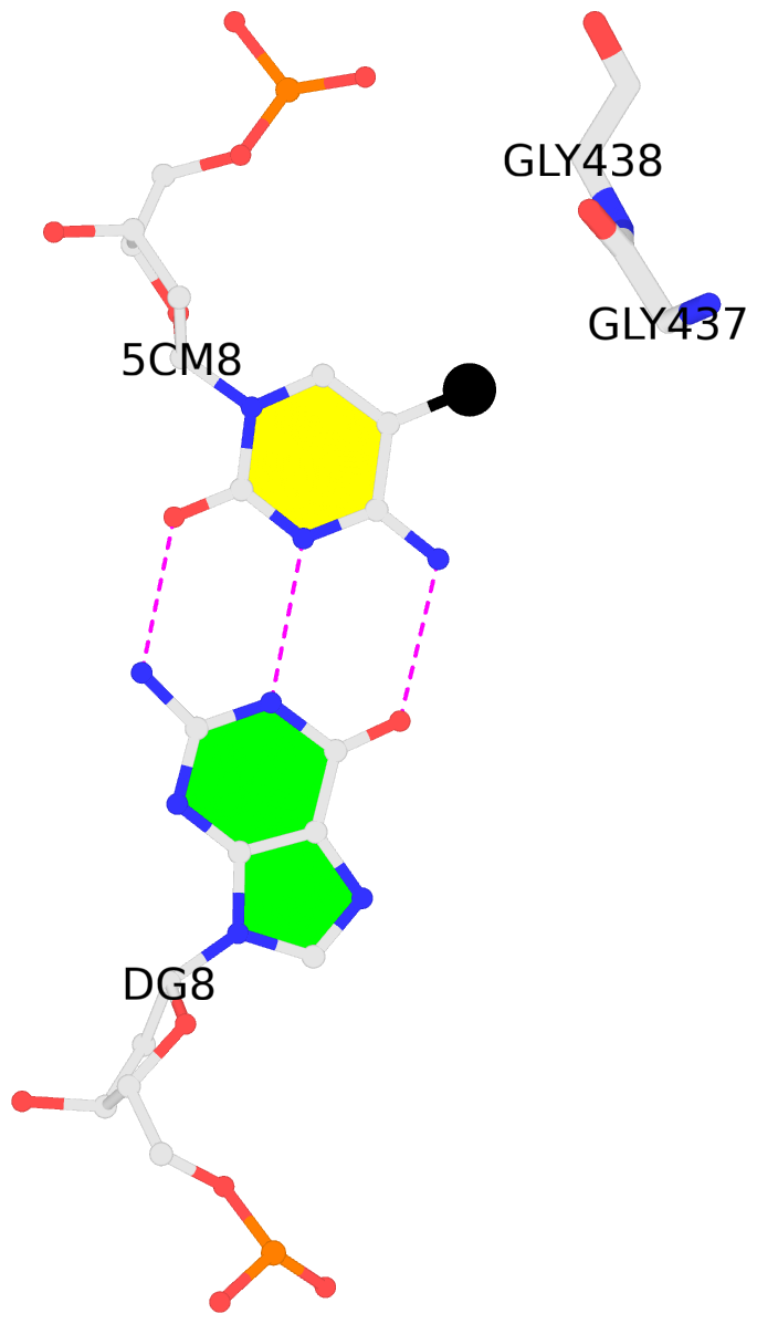 |
No. 1 I.5CM8: download PDB file
for the 5mC entry
hydrophobic-with-A.GLY437 hydrophobic-with-A.GLY438 is-WC-paired is-in-duplex [+]:TcT/AGA
|
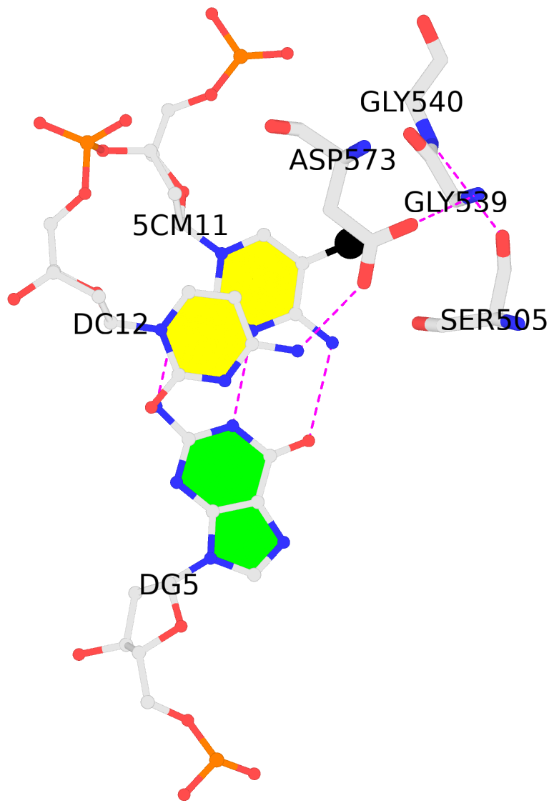 |
No. 2 I.5CM11: download PDB file
for the 5mC entry
stacking-with-A.ASP573 is-WC-paired is-in-duplex [+]:AcC/GGT
|
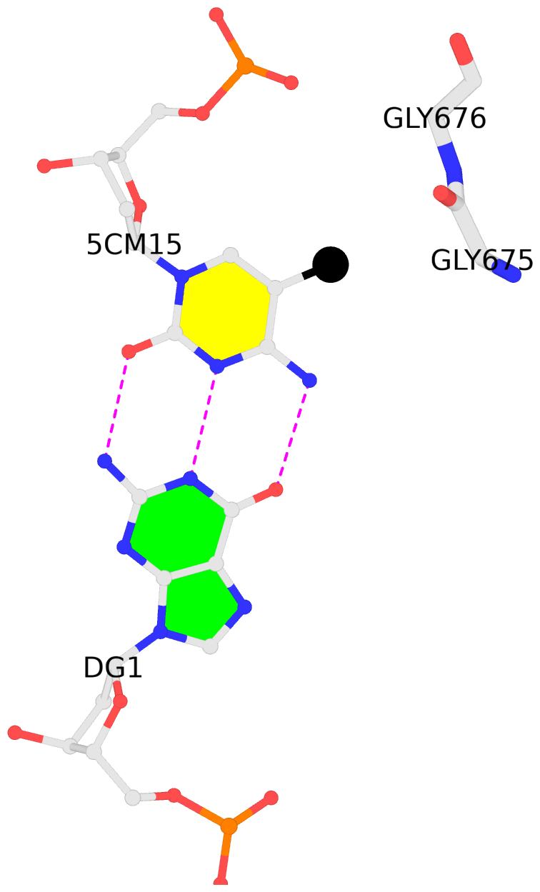 |
No. 3 I.5CM15: download PDB file
for the 5mC entry
hydrophobic-with-A.GLY675 hydrophobic-with-A.GLY676 is-WC-paired is-in-duplex [+]:CcC/GGG
|
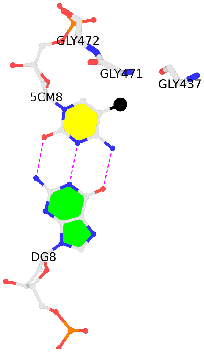 |
No. 4 G.5CM8: download PDB file
for the 5mC entry
hydrophobic-with-B.GLY437 hydrophobic-with-B.GLY471 hydrophobic-with-B.GLY472 is-WC-paired is-in-duplex [+]:TcT/AGA
|
 |
No. 5 G.5CM11: download PDB file
for the 5mC entry
stacking-with-B.ASP573 is-WC-paired is-in-duplex [+]:AcC/GGT
|
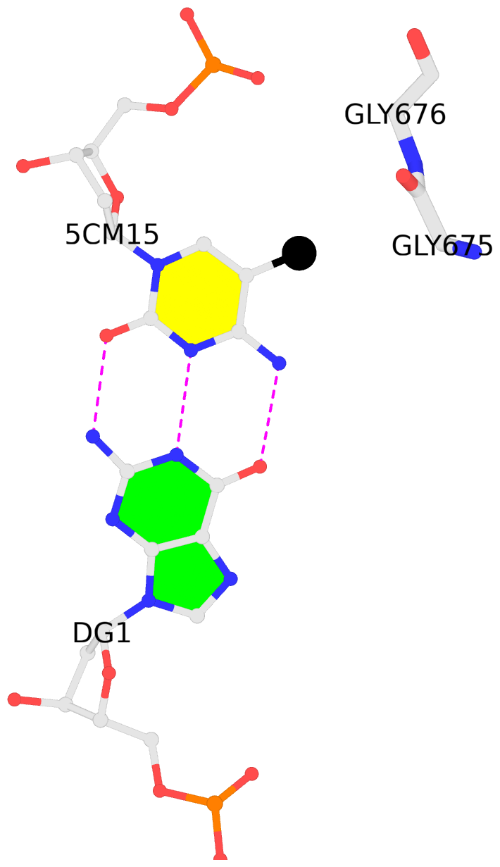 |
No. 6 G.5CM15: download PDB file
for the 5mC entry
hydrophobic-with-B.GLY675 hydrophobic-with-B.GLY676 is-WC-paired is-in-duplex [+]:CcC/GGG
|